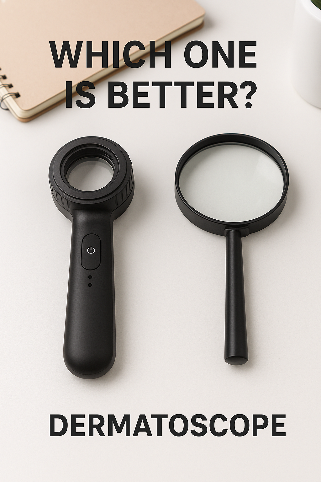
Dermatoscope vs Magnifying Glass: Which Tool Is Better for Skin Examination?
This guide breaks down the differences in magnification, diagnostic accuracy, lighting, use cases, and overall performance—so you can understand which tool is best suited for reliable skin evaluation. Dermatoscope vs Magnifying Glass
3 min read
Examining the skin closely can reveal early signs of melanoma, changes in moles, or other dermatological conditions. Whether you are a clinician, a medical student, or someone monitoring your own skin health, you may wonder how a dermatoscope compares to a magnifying glass. Both enlarge the skin’s surface, but they serve very different purposes and offer dramatically different results.
This guide breaks down the differences in magnification, diagnostic accuracy, lighting, use cases, and overall performance—so you can understand which tool is best suited for reliable skin evaluation.
What Is a Magnifying Glass?
A magnifying glass is a simple optical tool commonly used for reading fine print, examining objects, or performing general household tasks. It consists of a single convex lens that enlarges whatever is placed beneath it.
How a Magnifying Glass Works
Uses ambient light
Provides low-level magnification
Offers a limited field of view
Often introduces optical distortion
Magnifying glasses aren’t designed for medical evaluations. They can help you look at the surface of the skin, but they cannot show deeper structures or subtle changes that indicate early pathology.
What Is a Dermatoscope?
A dermatoscope is a medical diagnostic tool specifically engineered for examining skin lesions, pigmented spots, rashes, and suspicious moles. It is commonly used in dermatology clinics and increasingly in primary care settings.
Key Features of a Dermatoscope
Advanced optical system designed for clarity and distortion-free magnification
Built-in LED illumination (polarized and non-polarized)
10x medical-grade magnification
Contact or non-contact plates to stabilize the view
Ability to reveal subsurface skin structures invisible to the naked eye
A dermatoscope is an essential device for detecting melanoma and differentiating between benign and malignant lesions with far greater accuracy than a visual exam alone.
How Each Tool Works
Optical Differences
A magnifying glass provides basic magnification but struggles with:
Edge blurring
Chromatic aberration
Poor depth visualization
In contrast, a dermatoscope uses:
Multiple lens elements
Anti-reflective coatings
A wider field of view
High-resolution optics
This results in a sharper, more accurate image with consistent clarity across the entire lens.
Illumination Differences
Illumination is one of the most important aspects of skin imaging.
Magnifying Glass
Relies on available room light
Prone to glare and reflection
Cannot penetrate surface texture
Dermatoscope
Uses LED lighting designed for skin evaluation
Offers polarized light to suppress the skin’s surface reflection
Reveals pigment networks, vascular structures, and subsurface morphology
Can switch to non-polarized light to view surface features
This difference alone makes the dermatoscope a dramatically superior diagnostic tool.
Diagnostic Capabilities Compared
Surface vs. Subsurface Visualization
A magnifying glass lets you look only at surface-level changes—useful for examining:
Dryness
Scaling
Visible borders
A dermatoscope, however, enables visualization of:
Pigment networks
Dots and globules
Vascular patterns
Regression structures
Architectural disorder
These features are foundational in modern dermoscopy and essential for detecting melanoma early.
Accuracy and Reliability
Studies consistently show that dermatoscopy significantly increases:
Melanoma detection sensitivity
Diagnostic accuracy
Clinician confidence
A magnifying glass provides no improvement over the naked eye in diagnosing skin cancers.
Use Cases: When Each Tool Is Appropriate
When a Magnifying Glass Is Helpful
Casual self-checks
Viewing simple skin details
Looking at insect bites or splinters
Quick household observations
It is not suited for medical evaluation.
When a Dermatoscope Is Necessary
Clinical skin examinations
Diagnosing pigmented lesions
Melanoma screening
Monitoring mole changes
Teledermatology or smartphone imaging
Training medical students
Dermatoscopes are used worldwide because they allow early identification of concerning lesions that often look normal to the naked eye—or through a magnifying glass.
Pros and Cons of Each Tool
Magnifying Glass
Pros
Low cost
Easy to use
Widely available
Cons
No illumination
Limited magnification
No polarization
High distortion
Not diagnostic
Dermatoscope
Pros
Medical-grade image quality
Polarized and non-polarized lighting
Subsurface visualization
Wider field of view
Proven diagnostic accuracy
Cons
More expensive
Requires some training
Needs cleaning or sterilization after contact
Safety and Hygiene Considerations
A magnifying glass poses no medical safety risk, but it also provides no diagnostic benefit.
Dermatoscopes, especially contact models, must be:
Disinfected between uses
Used with disposable contact plates when possible
Handled with care in clinical settings
Non-contact dermatoscopy is also available for patients with sensitive or infectious lesions.
Cost Comparison
Magnifying glasses are inexpensive tools designed for general use.
Dermatoscopes cost more because they contain:
Precision optical systems
High-quality LEDs
Polarizing filters
Durable, medical-grade components
The investment reflects their professional diagnostic capability and essential role in modern dermatology.
Expert Recommendations
Dermatologists overwhelmingly recommend dermatoscopy as the standard method for evaluating suspicious skin lesions. Major medical organizations support its use because it:
Improves melanoma detection rates
Reduces unnecessary biopsies
Provides better documentation and monitoring
Enhances diagnostic confidence
A magnifying glass simply cannot replace a dermatoscope in any clinical scenario.
Conclusion: Dermatoscope vs Magnifying Glass
While both tools enlarge the skin, their roles are fundamentally different.
A magnifying glass is suitable for casual observation, but it cannot reveal diagnostic details or subsurface structures. It is not a medical instrument and should not be relied upon for screening skin lesions.
A dermatoscope offers superior magnification, advanced illumination, and the ability to visualize the deeper architecture of the skin—making it the clear choice for clinicians and anyone serious about accurate skin examination.
If your goal is reliable skin assessment, whether professional or personal with guidance from a dermatologist, the dermatoscope is the tool you need.
Stay Updated, Latest News
This website contains affiliate links, and we may earn a commission at no extra cost to you.